
︎
COLORANT KEY
To determine dichotomous UVR and IR responses, please refer to the L* value of your sample from the CIELAB color space.
DARK: L* ≤ 80
BRIGHT: L* ≥ 81
If reference value: X-rite Color Checker Classic square A4: VIS: L*94, IR: L*96, UVR: L*35

STEP 1. Open your calibrated images in photoshop - UVR & IR.
* For best results, create a tonal adjustment curve profile for each band and apply to all files using as many Spectralon you have available. VIS, IR, and UVR should all match L* in Spectralon values (+/- 3), here is a visual reference for VIS, IR, UVR:
STEP 2. *Optional step* File > Scripts > Load Files into Stack > Add Open Files + Attempt to Automatically Align Source Images.
STEP 3. Select Color Sampler tool, sample size: 31 by 31 average
STEP 4. Click on the area that you want to sample, you will see the reading pop up. Make sure you change the color reading to LAB by clicking on the eyedropper dropdown arrow in the INFO panel underneath the #1. If you stacked the files, you can now toggle layer visibility to view readings from each layer. You can see in the reading below, that the lightness (L*) value reads 60, which is ≤ 80, meaning the sample is ‘DARK’.

︎
COLORANT KEY + MUNSELL CALCULATOR
FLUORESCENCE: CHROMA > 20
[If less than 20, UVF: NONE]
If reference value: UV Innovations Target Low, brightest L*= 55 a*=0 b*=0

To determine UVF response, please use the MUNSELL calculator to convert Lab values to Munsell Hue and Chroma.
*The system does not provide quantitative fluorescence intensity in physical units (e.g., radiance or lumens). If precise fluorescence intensity measurements are required, consider using spectrophotometers, fluorometers, or imaging systems with calibrated fluorescence intensity scales. These tools can provide absolute values rather than relying on visual assessments.
STEP 1. Open your calibrated image in photoshop - UVF.
** Double check that your reference value (UV Innovations Target) is balanced. Gray values a* b* = 0 or as close as possible on each image.
For more info, see their website for workflow. As many camera RAW programs have a white balance setting that maxes out at 50,000 Temp. You may need to do some color grading to get a properly balanced target OR choose a standard and follow it.
STEP 2. Select Color Sampler tool, sample size: 31 by 31 average
STEP 3. Click on the area that you want to sample, you will see the reading pop up. Make sure you change the color reading to LAB by clicking on the eyedropper dropdown arrow in the INFO panel underneath the #1.
STEP 4. Enter your LAB values into the Munsell Calculator to convert.
Munsell color notation: HUE, VALUE/CHROMA
UVF HUE: R, YR, Y, GY, G, BG, B, PB, P, and RP
[Hue has been divided into the 10 Munsell hue sectors, Red, Yellow-Red etc.]

︎
VECTOR CHART
To calculate a response vector graph, please use the VECTOR CHART calculator to chart Lab values measured from each wavelength band.
*The calculator only provides discrete, approximate values. For more accurate and continuous values, please use a spectrophotometer.
Vector response chart for Cochineal ︎
STEP 1. Open your calibrated images in photoshop - UVR & VIS & IR.
** Double check that your reference value (X-rite Color Checker Classic A4: L*=96) on each image.
STEP 2. Separate VIS file into the three channels, R + G + B
STEP 3. Optional step* File > Scripts > Load Files into Stack > Add Open Files + Attempt to Automatically Align Source Images.
STEP 4. Select Color Sampler tool, sample size: 31 by 31 average
STEP 5. Click on the area that you want to sample, you will see the reading pop up. Make sure you change the color reading to LAB by clicking on the eyedropper dropdown arrow in the INFO panel underneath the #1.
STEP 6. Enter your L* values into the Vector Chart Calculator to generate chart.
︎
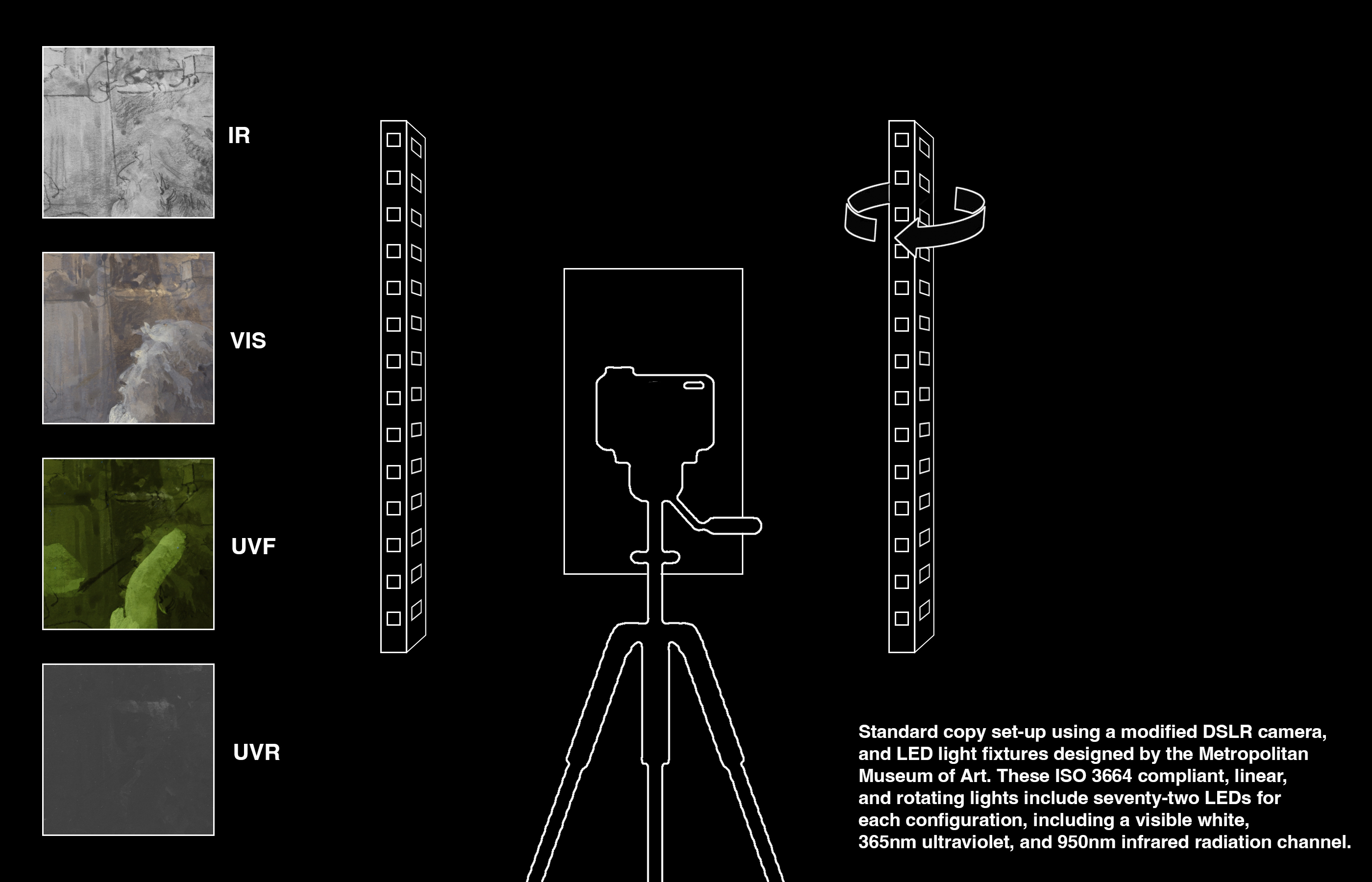
Imaging set - up:
Camera: Modified Canon 5DSR full spectrum conversion.
Lens: Coastal Optics Apochromatic lens, 60mm, 1:4.
Filters: VIS - IDAS-UIBAR III [400-680nm], UVR - XNite 330C + XNite BP1 [270-400nm], UVF - IDAS-UIBAR III + Wratten 2E [420-680nm], IR - XNite 830 [830-1100nm].
Lights: LED bulbs, standard copy set-up using lights designed by the Metropolitan Museum of Art: These 24” linear fixtures were designed to provide consistent uniform illumination for technical cultural heritage imaging. D50 98CRI ISO 3664 compliant visible white channel, 365nm UV, and 950nm IR. The unique compact form-factor incorporates a rotating body to maintain consistent lighting geometry for critical comparative study and multi-band imaging techniques.
Targets: x-rite classic mini color checker, UV innovations target [low/medium], labsphere spectralons.
Backgound paper: Color-aid Gray 4 (non-fluorescing neutral gray).
Questions: lauramargaret.ramsey(at)metmuseum.org
*IDAS-UIBAR III no longer available.
Comparable options, in order of performance1
VIS: MidOpt BP550 [390-740], UVF: MidOpt BP550 + Wratten 2E [420-740]
VIS: MidOpt SP700 [400-780], UVF: MidOpt SP700 + Wratten 2E [420-780]
VIS: PECA 918 [380-740], UVF: PECA 918 + Wratten 2E [420-740]
1. Mastandrea, W.J., K. McFarlin, C. Heins, A. Grant, R. Greenberg, A. Serotta, and D. Kriss. “The 12th International Round Table on Polychromy in Ancient Sculpture and Architecture.” In Letting the Light In: A Comparative Assessment of Visible Bandpass Filters Used in Multiband Imaging of Material Culture. Los Angeles, 2024.
︎
Colorants included as references: Kremer Pigment Register, 2002
︎
Additional References:
Cosentino, Antonino. “Identification of Pigments by Multispectral Imaging; a Flowchart Method.” Heritage Science 2, no. 1 (March 17, 2014). https://doi.org/10.1186/2050-7445-2-8.
Dyer, J., Verri, G., & Cupitt, J. Multispectral Imaging in Reflectance and Photoinduced Luminescence modes: A User Manual, 2013. Retrieved from https://www.britishmuseum.org/pdf/charisma-multispectral-imaging-manual-2013.pdf
Fiske, Betty, and Linda Stiber Morenus. “The Book & Paper Group Annual, v. 23 (2004)” 23 (2004): 21–32.
Gettens, Rutherford J., and George L. Stout. Painting materials: A short encyclopaedia. New York: Dover Publications, 2015.
Mastandrea, W.J., K. McFarlin, C. Heins, A. Grant, R. Greenberg, A. Serotta, and D. Kriss. “The 12th International Round Table on Polychromy in Ancient Sculpture and Architecture.” In Letting the Light In: A Comparative Assessment of Visible Bandpass Filters Used in Multiband Imaging of Material Culture. Los Angeles, 2024.
McGlinchey Sexton, J., Messier, P., & Chen, J. J. Development and Testing of a Fluorescence Standard for Documenting Ultraviolet Induced Visible Fluorescence. In 42nd Annual American Institute for Conservation Meeting (pp. 1–42). San Francisco: American Institute for Conservation, 2014.
Measday, Danielle, Charlotte Walker , and Briony Pemberton. A Summary of Ultra-Violet Fluorescent Materials Relevant to Conservation , n.d. https://primastoria.wordpress.com/wp-content/uploads/2017/03/uv-relevant-to-conservation.pdf.
Stuart, Barbara. Analytical Techniques in Materials Conservation. Chichester, England: John Wiley & Sons, 2010.
COLORANT KEY & FIELD GUIDE
L. M. Ramsey, 2024